While it hasn’t been long since our latest AutoContour release, we’ve been diligently working on the latest additions to make contouring even better. As always, we’ve listened to your feedback and incorporated your suggestions. In this latest batch release, we include dozens of new structure models to add to our existing offerings, including additional MR models, vertebral bodies, and a number of variations to existing models to conform to your department’s unique preferences.
AutoContour is not available in all markets.
MR Models
With the announcement of v2.2, we released our first MR models, a limited batch of structures for cranial contouring. We’ve heard your requests for more, and we’re excited to say we have added to our list of available MR models.
Our MR T1-post model list continues to grow following guidelines by the EPTN Consensus Neuro-Oncology Atlas, adding the following to the available list of structure models:
- OpticTract (Left and Right)
- Eye (Left and Right)
- Pituitary
- Hypothalamus
- Hypo_True (a variation of the hypothalamus that does not include mammillary bodies)
- Cerebellum
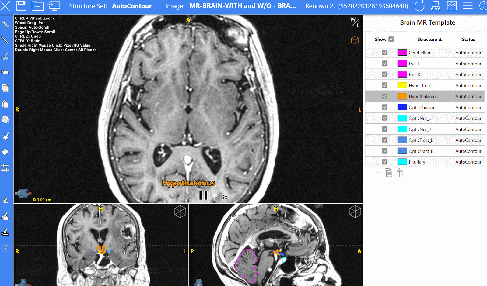 New cranial MR models were added to the initial offering, expanding capabilities for SRS and SRT treatment regimens.
To complement our MR T1 post models, we’ve created MR T2 models for prostate anatomy. The models we’ve created for the prostate, prostate gland, and seminal vesicles utilize T2, small FOV datasets, which are oft-associated with these treatments.
New for prostate case contouring using T2 scan data:
- Prostate
- Prostate Gland
- Seminal Vesicles
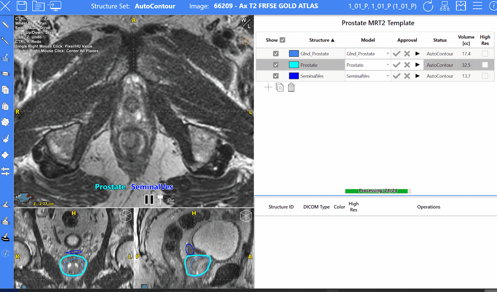
Scroll-through image of new prostate MR anatomy featuring the prostate, prostate gland, and seminal vesicles.

Vertebral Bodies
In the latest release, a variety of vertebral body models are now available. In addition to providing a full vertebral column model that contours all vertebral bodies together, we have individual vertebral bodies for each and every cerebral, thoracic, and lumbar segment you may need. These models can be helpful in quickly identifying vertebral anatomy and ensuring the correct location of other contours.
For example, having L1 and L2 contoured will help users to quickly locate where the spinal cord should end and where the cauda equina should begin.
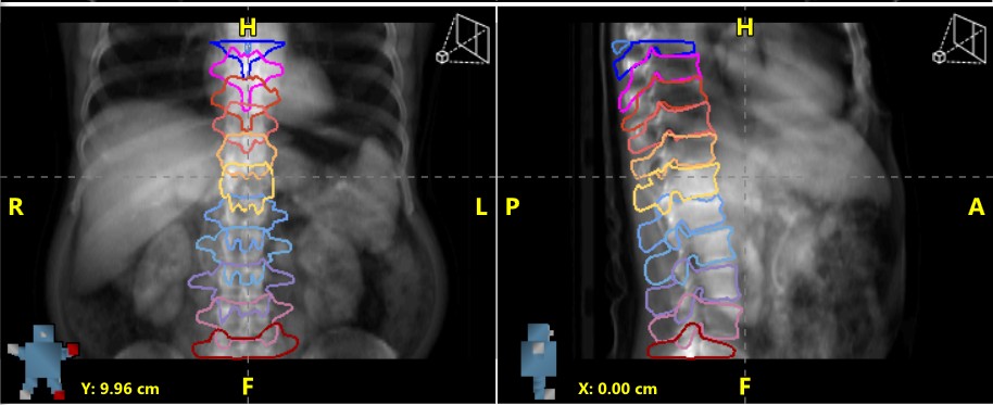
AutoContour results for individual vertebral bodies.
New Model Variations
Radformation is aware that clinical needs vary. This is especially true when it comes to structure contouring. Even if departments follow consensus guidelines, multiple publications for various anatomical regions exhibit subtle differences for each structure. AutoContour now better accommodates these unique needs with new models for multiple structures based on various sets of guidelines.
This batch release of new structure models includes new variations to existing models. Users can now choose the model that best suits their clinical preferences. We've made room in our growing model library for the following structures:
- Bowel
- Larynx
- Axial Lymph Nodes
- Internal Mammary Lymph Nodes (IMNs)
- Supraclavicular Lymph Nodes
- Chestwall
- Bladder
- Rectum
Bowel Model Variations
New bowel models have been added for flexibility so that users can use the model that best fits their clinic.
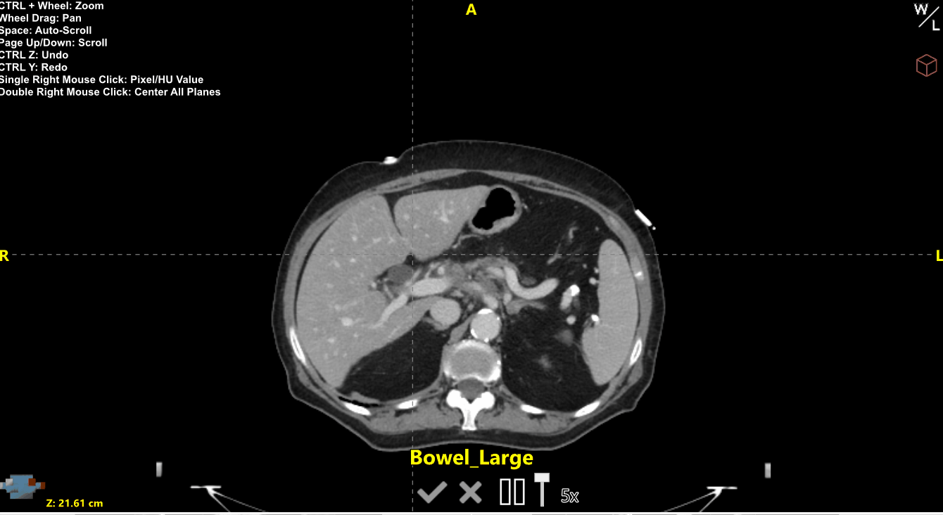
Our Bowel Bag model has been updated to avoid over-contouring into bone or muscle, and can be used when a generous contour of all the bowel space is needed. Our new Bowel model includes all bowel loops but no bowel space. The Small Bowel model only includes loops of the small bowel—with no portions of the colon—and includes the duodenum. The Large Bowel model includes all portions of the colon, including the sigmoid colon. Duodenum and Sigmoid Colon models will also be available separately from these bowel models.
AutoContour results for Large Bowel (Yellow) and Small Bowel (Red) for a scan with contrast in the bowel.
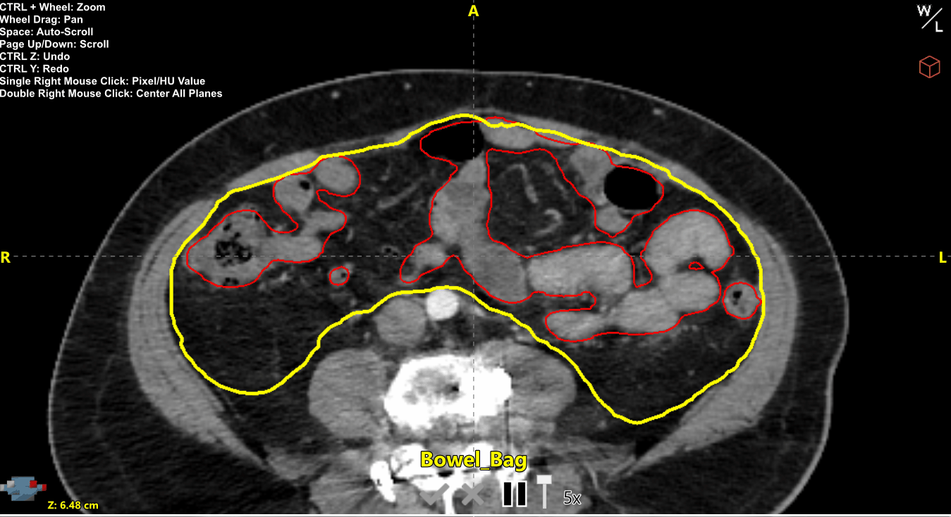
AutoContour results for Bowel Bag (Yellow) and Bowel (Red) for a scan without contrast in the bowel.
Larynx Model Variations
Our new larynx models were designed to contour the Glottic Larynx (Larynx_Glottic) and the Supraglottic Larynx (Larynx_SG) separately but also a combination of these regions (Larynx_NRG). These new models have different cranial and caudal borders but are also trained to avoid the air in these regions, which differs from the previously released Larynx model.
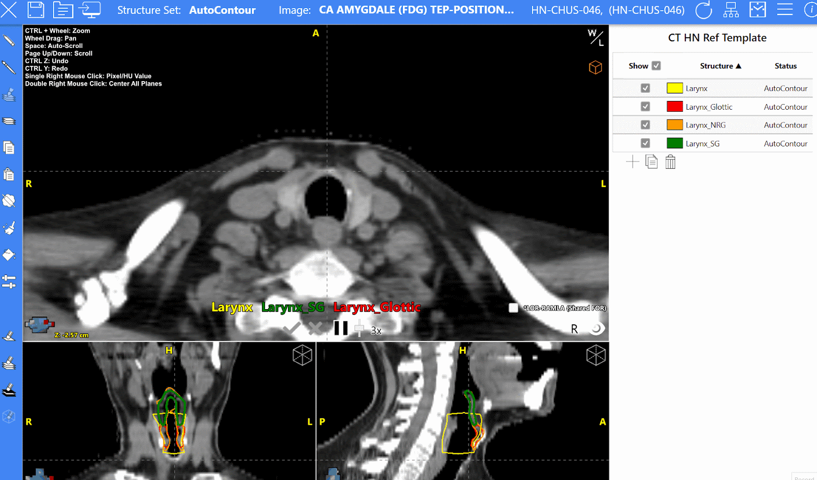
AutoContour results for Larynx (Yellow), Supraglottic Larynx (Green), Glottic Larynx (Red), and Larynx_NRG (Orange) on a head and neck scan.
Breast Node Model Variations
New models following RADCOMP guidelines for breast radiotherapy are available for the departments that use them. These models are denoted by an RC (e.g., IMN_RC_L/R, Sclav_RC_L/R, and Chestwall_RC_L/R). The differences between the IMN and Supraclavicular Nodes contouring guidelines are shown here:


AutoContour results for IMN (Yellow) and RADCOMP IMN (Red).
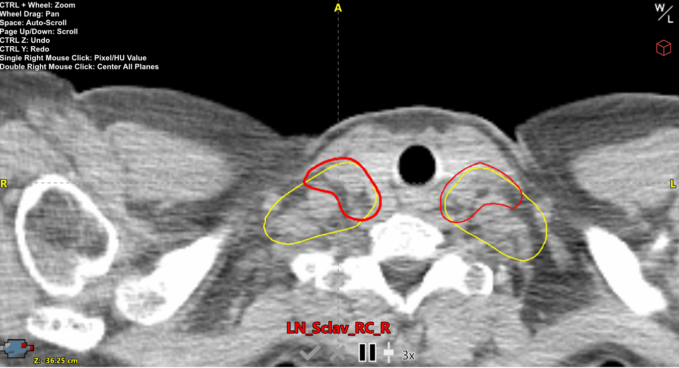
AutoContour results for Supraclavicular Nodes (Yellow) and RADCOMP Supraclavicular Nodes (Red).
Along with these new RADCOMP breast nodes, we are excited to release individual Axilla Node models following The RTOG Breast Cancer Consensus Guidelines, as well as a combined Level II and III model, as shown below:
AutoContour results for the new split Axilla nodes, Axilla Level 1 (Orange), Axilla Level 2 (Yellow), and Axilla Level 3 (Red)
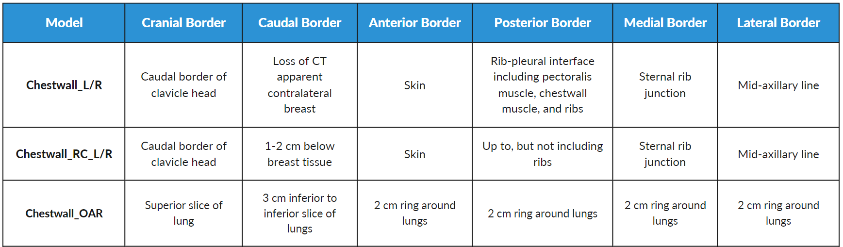
Chestwall Model Variations:
Our new chestwall model variations differ in clinically relevant ways. There are now two chestwall structure models, a standard, existing Chestwall structure and another that conforms to RADCOMP border guidelines. Though these are similar structures (and even share some border definitions), they are separate, unique models.
A new Chestwall OAR was designed to be used for lung cases—where the chestwall is contoured primarily as an organ at risk versus a target—while Chestwall (regular and RADCOMP) structures are intended to be used for breast cases (specifically, mastectomy cases). Because of the inherently different needs for lung cases versus breast cases, the Chestwall and Chestwall OAR structures do not closely resemble each other.
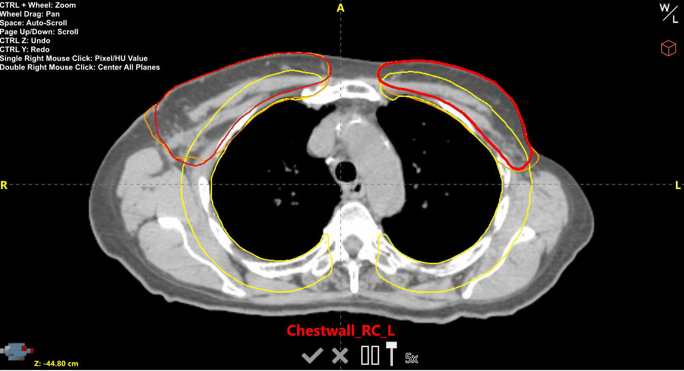
AutoContour results for Chestwall left and right (Orange), RADCOMP Chestwall left and right (Red), and OAR Chestwall (Yellow).
Bladder and Rectum Model Variations
In our own internal tests—as well as from clinical feedback—we’ve come to realize that Bladder and Rectum structure contours are especially affected by local anatomy differences between male and female patients. Our original Bladder and Rectum models do not perform as well on female patients due to several factors, such as the presence of the uterus. We have now trained new female models to account for these factors, dubbing them Bladder_F and Rectum_F. These models also exhibit improved performance on HDR cases, since they were trained with these in mind.
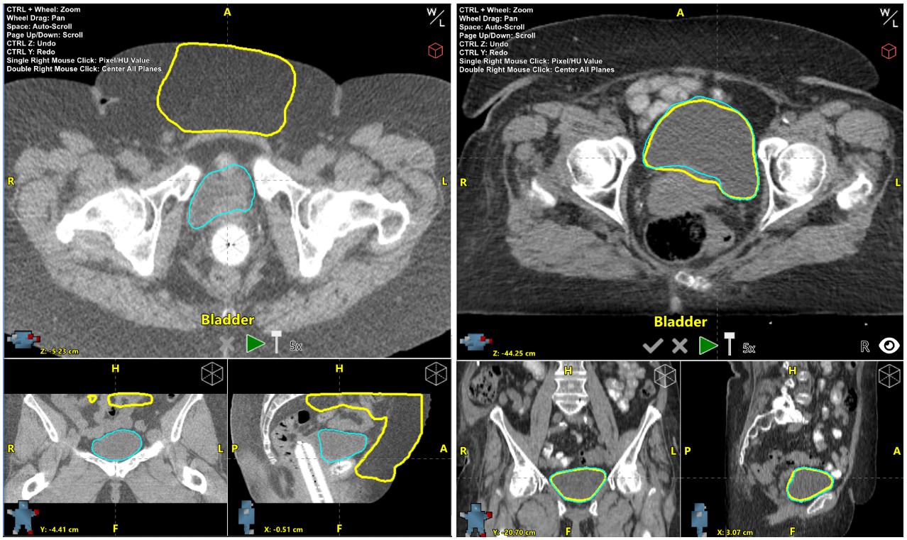 Left: Bladder (Yellow) AutoContour results compared to Bladder_F (Cyan) AutoContour results for a female pelvis HDR scan. Right: Bladder (Yellow) AutoContour results compared to Bladder_F (Cyan) Autocontoured results for a female pelvis scan.
Benefits and Limitations to New Models
Having a selection of unique models to complement existing offerings is beneficial as it provides users with multiple options when one model may not be suitable for a department's protocols or even for a particular scan. AI-based contouring is an incredible technology that has transformed the pre-treatment workflow, but it's worth noting that AI-based algorithms do have some limitations and may not always perform optimally for some scans.
In the abdomen, for example, the distinction between the small and large bowel can be challenging to discern in some regions, even for an experienced dosimetrist. Our models perform well and provide reliable options for bowel contouring, but it's possible to see some level of overlapping contours for the small and large bowel. In addition, as with any AI-based segmentation algorithm, significant changes in contrast within a scan can affect the segmentation results. Still, our models are helpful in reducing the number of necessary edits, even in cases where contrast or artifacts are present. Our new model variations are designed to provide additional choices for users in such scenarios.
Conclusion
In conclusion, Radformation continues to push the boundaries of AI autosegmentation with AutoContour’s latest structure release. With the addition of new cranial and prostate MR models, clinicians now have even more options for precise and efficient contouring. The inclusion of individual vertebral body models and new model variations further enhances the flexibility and customization of contouring based on clinical preferences.
While there may be limitations with any AI-based algorithm, these new models are designed to reduce the number of required edits and improve contouring accuracy. Radformation remains committed to listening to user feedback and continuously improving AutoContour to provide the best contouring solutions for radiation oncology departments. So with this latest AutoContour structure release, Radformation continues to drive forward, empowering clinicians with advanced tools for efficient and accurate contouring in radiotherapy planning.

Leave a comment